Animal cell under microscope information
Home » Trending » Animal cell under microscope informationYour Animal cell under microscope images are available. Animal cell under microscope are a topic that is being searched for and liked by netizens today. You can Download the Animal cell under microscope files here. Find and Download all free photos and vectors.
If you’re searching for animal cell under microscope images information related to the animal cell under microscope topic, you have come to the right blog. Our website always gives you suggestions for downloading the maximum quality video and image content, please kindly search and locate more informative video articles and images that match your interests.
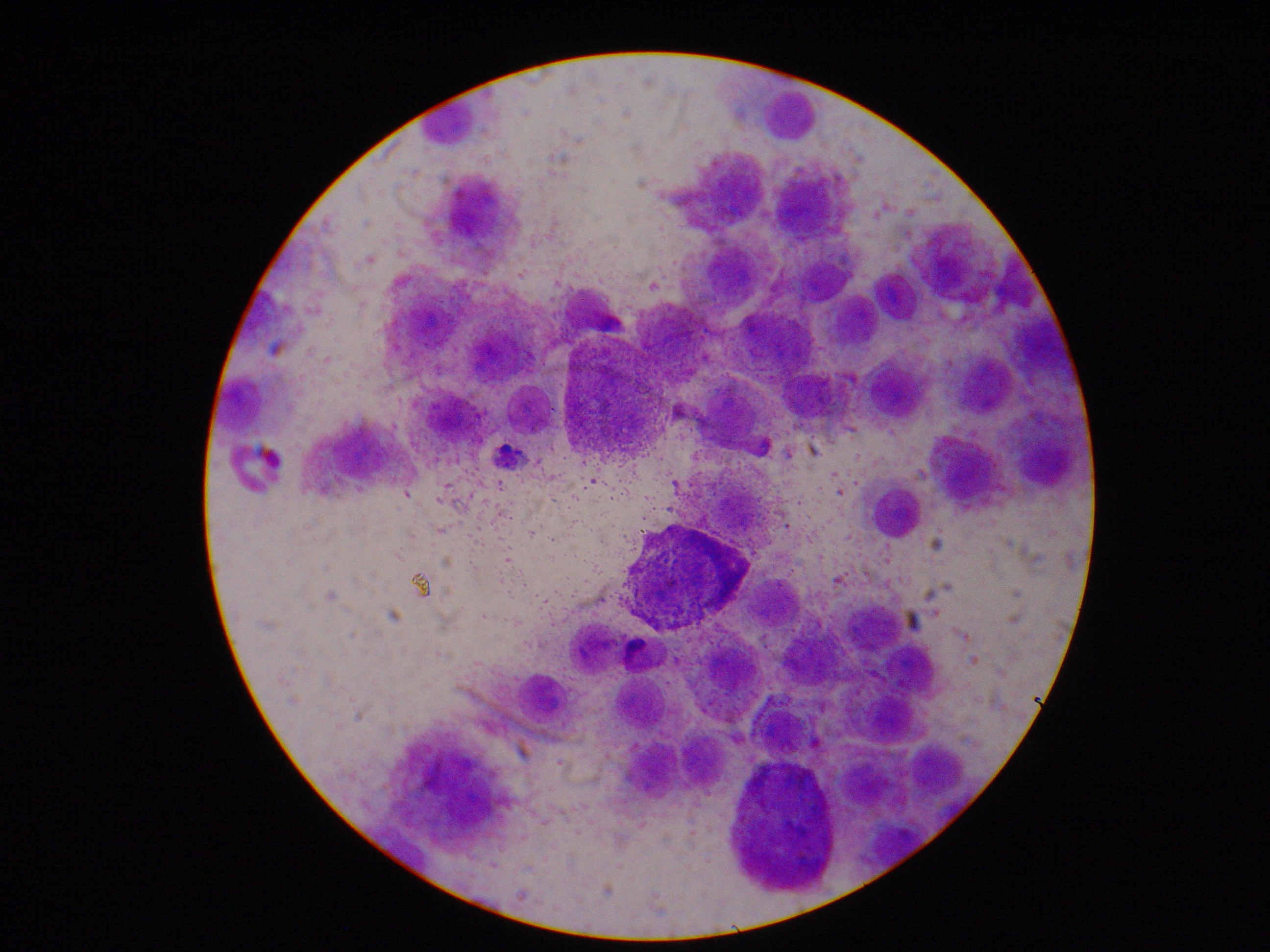
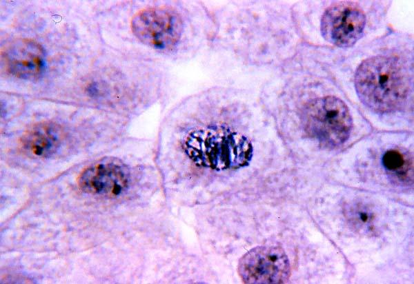
Animal Cell Under Microscope. Find and view animal cells using a microscope. Below the basic structure is shown in the same animal cell, on the left viewed with the. An animal cell represents an eukaryotic cell in which true nucleus and other membrane bound organelles such as mitochondria. But at the same time it is interpretive.
 Mast Cell Tumors in Dogs A Common Canine Skin Cancer From criticalcaredvm.com
Diagram of animal cell under electron microscope. The diagram is very clear and labeled. There are one or more cells that form organism. Diagram of animal cell under electron microscope. It was not until good light microscopes became available in the early part of the nineteenth century that all plant and animal tissues were discovered to be aggregates of individual cells. Find and view animal cells using a microscope.
Diagram of animal cell under electron microscope.
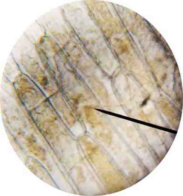
Most cells, both animal and plant, range in size between 1 and 100 micrometers and are thus visible only with the aid of a microscope. Royalty free stock photos a typical cell labeled cell diagram animal cell cells worksheet. See animal cell under microscope stock video clips. But at the same time it is interpretive. Epithelial cells surround the internal surface of the mouth which can be taken out using finger nails or a small spoon. Animal cell diagram under electron microscope.
 Source: youtube.com
Most of the cells are microscopic in size and can only be seen under the microscope. Contents 1 when looking at plant and animal cells with an electron microscope you notice that the plant cells. Magnification = size of image / size of real object structure of animal cell and plant cell under microscope + diagrams learn the structure of animal cell and plant cell under light microscope. Below the basic structure is shown in the same animal cell, on the left viewed with the. Find and view animal cells using a microscope.
 Source: perkinselearning.org
Magnification = size of image / size of real object structure of animal cell and plant cell under microscope + diagrams learn the structure of animal cell and plant cell under light microscope. Find and view animal cells using a microscope. Structures viewed under an optical microscope can be measured using the formula: What type of microscope is needed to see a plant cell? Contents 1 when looking at plant and animal cells with an electron microscope you notice that the plant cells.
 Source: easynotecards.com
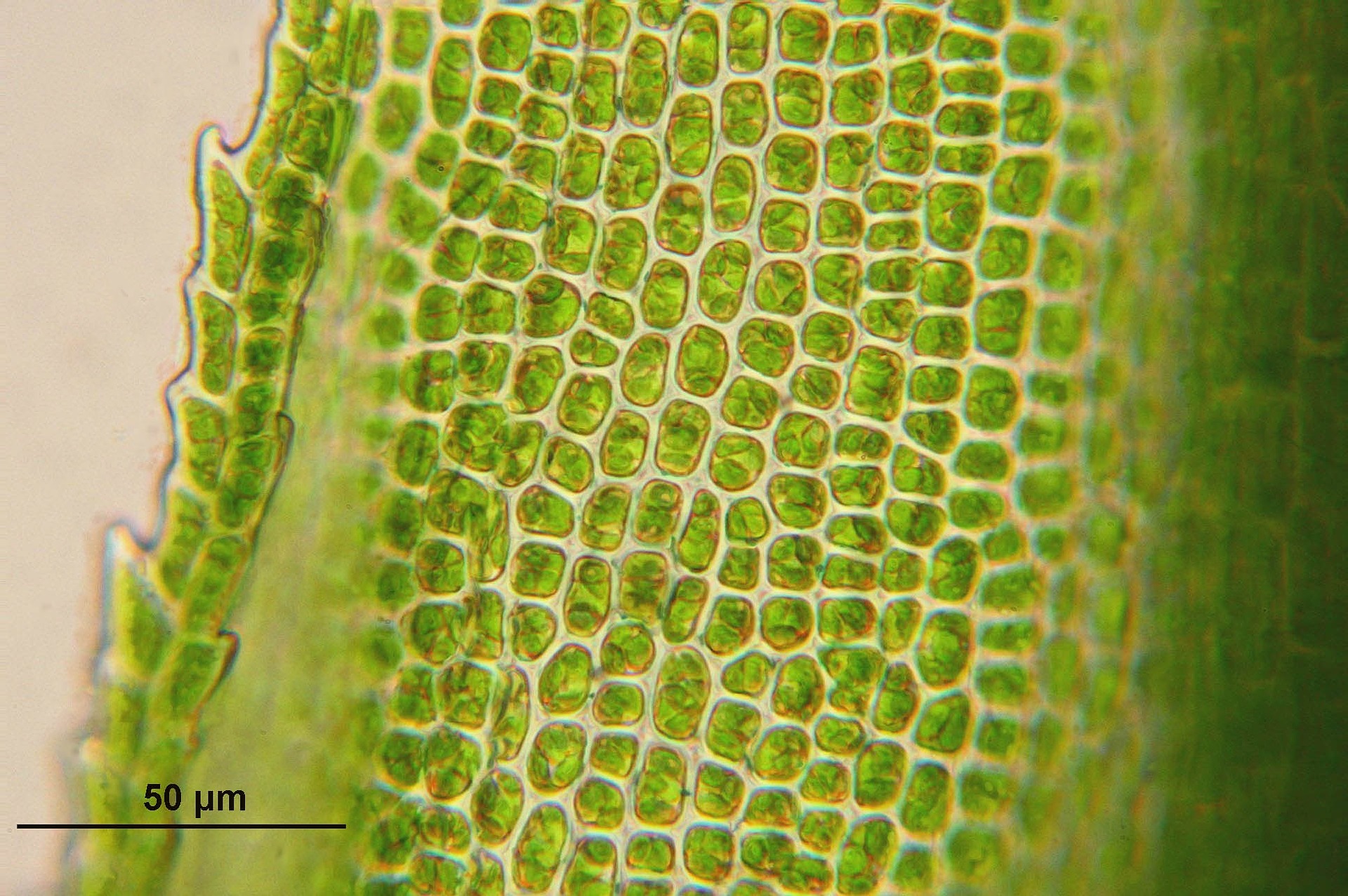
Cell 8 pictures of plant cells under a microscope plant cell structure under microscope plant and animal cells plant cell structure plant cell. So it is important to note that. Compare an animal cell to a plant cell. Magnification = size of image / size of real object structure of animal cell and plant cell under microscope + diagrams learn the structure of animal cell and plant cell under light microscope. Browse 4,547 animal cells under microscope stock photos and images available or start a new search to explore more stock photos and images.
 Source: scienceprofonline.com
Nucleus, cytoplasm, cell membrane, chloroplasts and cell wall (last 2 organelles are only present in plant cells). Animal cells almost all animals and plants are made up of cells. Use the equation m=i/a to. What parts of an animal cell can you see under a light microscope? You know, animal cell structure contains only 11 parts out of the 13 parts you saw in the plant cell diagram, because chloroplast and cell wall are available only in a plant cell.
 Source: criticalcaredvm.com
Angelo on august 20, 2021. Use the equation m=i/a to. Epithelial cells surround the internal surface of the mouth which can be taken out using finger nails or a small spoon. Meiosis cell division 3d cell cellular division embryo 3d cell animal embryo reproductive health blood cells under microscope the cell cytyoplasm cytoplasm. Angelo on august 20, 2021.
This site is an open community for users to share their favorite wallpapers on the internet, all images or pictures in this website are for personal wallpaper use only, it is stricly prohibited to use this wallpaper for commercial purposes, if you are the author and find this image is shared without your permission, please kindly raise a DMCA report to Us.
If you find this site good, please support us by sharing this posts to your preference social media accounts like Facebook, Instagram and so on or you can also bookmark this blog page with the title animal cell under microscope by using Ctrl + D for devices a laptop with a Windows operating system or Command + D for laptops with an Apple operating system. If you use a smartphone, you can also use the drawer menu of the browser you are using. Whether it’s a Windows, Mac, iOS or Android operating system, you will still be able to bookmark this website.
Category
Related By Category
- 70s robot anime information
- Animated dd maps information
- Animal crossing new leaf mobile information
- Anime body base information
- Animal crossing jacobs ladder flower information
- Anime desserts information
- Animal paca information
- Animal crossing secrets information
- American animals review information
- Animal kingdom lodge rooms for 5 information