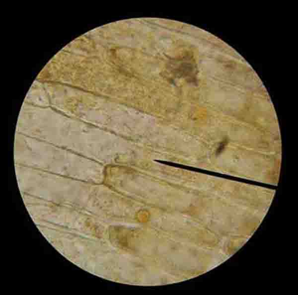
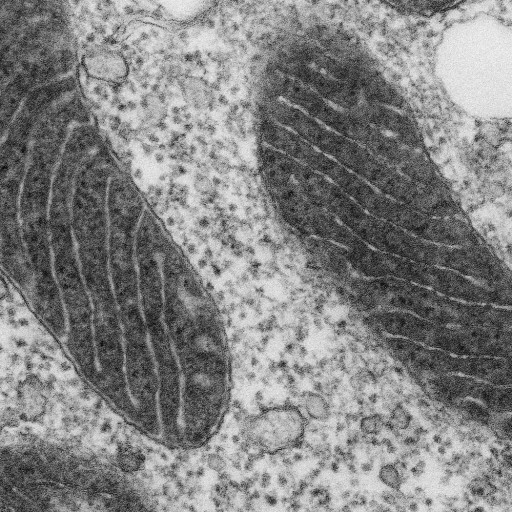
Animal cell under electron microscope information
Home » Trending » Animal cell under electron microscope informationYour Animal cell under electron microscope images are ready. Animal cell under electron microscope are a topic that is being searched for and liked by netizens now. You can Get the Animal cell under electron microscope files here. Get all free photos and vectors.
If you’re searching for animal cell under electron microscope pictures information related to the animal cell under electron microscope keyword, you have visit the right blog. Our website frequently provides you with suggestions for downloading the highest quality video and image content, please kindly hunt and find more enlightening video content and graphics that fit your interests.
Animal Cell Under Electron Microscope. How to get a lion as a. Animal cell diagram under electron microscope. We all keep in mind that the human body is quite elaborate and a method i found out to. Typical animal cell pinocytotic vesicle lysosome golgi vesicles golgi vesicles rough er (endoplasmic reticulum) smooth er (no ribosomes) cell (plasma) membrane mitochondrion golgi apparatus nucleolus nucleus centrioles (2) each composed of 9.
 Plankton May Hold Up Well to Ocean Acidification WIRED From wired.com
Microscope comes in different types that produce different result to see. Animal cell diagram under electron microscope. Eukaryotic is most complex cells consisting a true nucleus enclosed by a membrane. Below the basic structure is shown in the same animal cell, on the left viewed with the light microscope, and on the right with the transmission electron microscope. This is a colored scanning electron micrograph of human red and white blood cells: There is also another type of microscope called light microscope under a light microscope, the parts of a simple animal cell (e.g.
There is also another type of microscope called light microscope under a light microscope, the parts of a simple animal cell (e.g.
Angelo on november 24, 2021. Admin send an email 3 weeks ago. Animal cell diagram electron microscope. (a) identify cell structures (including organelles) of typical plant and animal cells from diagrams, photomicrographs and as seen under the light microscope using prepared slides and fresh material treated with an appropriate temporary staining technique: Compare an animal cell to a plant cell. That’s the major difference between plant and animal cells under microscope.
 Source: keele.ac.uk
Plant and animal cells have their similarities and differences. Generalized cell is used for structure of animal cell and plant cell to present the common parts, appearing in various parts of the bodies of animals and plants. The diagram is very clear and labeled. Observing plant cell or animal cell under microscope is important as a cell is a very small unit that can’t be seen with your naked eye. Schematic diagrams of typical animal and plant cells are shown in figure 1a and b.
 Source: wired.com
Generalized cell is used for structure of animal cell and plant cell to present the common parts, appearing in various parts of the bodies of animals and plants. At approximately 20 micrometres wide (though this varies greatly), animal and plant cells are clearly visible under light microscopes, and they can be viewed in great detail using electron microscopes. We have got 7 pic about diagram of animal cell seen under electron microscope images, photos, pictures, backgrounds, and more. See more articles in category: It was not until good light microscopes became available in the early part of the nineteenth century that all plant and animal tissues were discovered to be aggregates of individual cells.
 Source: columbia.edu
Cell 8 pictures of plant cells under a microscope plant cell structure under microscope plant and animal cells plant cell structure plant cell. There are two categories of cells, eukaryotic and prokaryotic. An electron micrograph of a typical plant cell is shown next. Light microscopes use lenses and light to magnify cell. Schematic diagrams of typical animal and plant cells are shown in figure 1a and b.
 Source: realclearscience.com
They are very tiny than what human eyes can see in general. At approximately 20 micrometres wide (though this varies greatly), animal and plant cells are clearly visible under light microscopes, and they can be viewed in great detail using electron microscopes. A typical animal cell (as seen in an electron microscope) medical images for powerpoint. In the given figure of an animal cell as observed under an electron microscope. Animal cell diagram under electron microscope.
 Source: br.pinterest.com
Generalized cell is used for structure of animal cell and plant cell to present the common parts, appearing in various parts of the bodies of animals and plants. It was not until good light microscopes became available in the early part of the nineteenth century that all plant and animal tissues were discovered to be aggregates of individual cells. Diagram 1.1 below shows an animal cell seen under the electron microscope. Generalized cell is used for structure of animal cell and plant cell to present the. Observing plant cell or animal cell under microscope is important as a cell is a very small unit that can’t be seen with your naked eye.
 Source: scienceprofonline.com
A typical animal cell (as seen in an electron microscope) medical images for powerpoint. Living cells cannot be observed using an electron microscope because samples are placed in a vacuum. Illustrate only a plant cell as seen under electron microscope. That’s the major difference between plant and animal cells under microscope. The electron microscope two main advantages high resolving power (short wavelength of electrons) as electrons negatively are charged the beam can be focused using electromagnets as electrons are absorbed by molecules of air, a.
 Source: kurzweilai.net
Compare an animal cell to a plant cell. A cell is the smallest functional and structural entity of life that it is easier observing animal cell under light microscope. These are both specific typesof cells, and from specific species. There are two categories of cells, eukaryotic and prokaryotic. Living cells cannot be observed using an electron microscope because samples are placed in a vacuum.
This site is an open community for users to do sharing their favorite wallpapers on the internet, all images or pictures in this website are for personal wallpaper use only, it is stricly prohibited to use this wallpaper for commercial purposes, if you are the author and find this image is shared without your permission, please kindly raise a DMCA report to Us.
If you find this site helpful, please support us by sharing this posts to your preference social media accounts like Facebook, Instagram and so on or you can also save this blog page with the title animal cell under electron microscope by using Ctrl + D for devices a laptop with a Windows operating system or Command + D for laptops with an Apple operating system. If you use a smartphone, you can also use the drawer menu of the browser you are using. Whether it’s a Windows, Mac, iOS or Android operating system, you will still be able to bookmark this website.
Category
Related By Category
- 70s robot anime information
- Animated dd maps information
- Animal crossing new leaf mobile information
- Anime body base information
- Animal crossing jacobs ladder flower information
- Anime desserts information
- Animal paca information
- Animal crossing secrets information
- American animals review information
- Animal kingdom lodge rooms for 5 information