Animal cell electron microscope labelled information
Home » Trend » Animal cell electron microscope labelled informationYour Animal cell electron microscope labelled images are available. Animal cell electron microscope labelled are a topic that is being searched for and liked by netizens now. You can Get the Animal cell electron microscope labelled files here. Get all royalty-free images.
If you’re looking for animal cell electron microscope labelled pictures information linked to the animal cell electron microscope labelled keyword, you have come to the ideal blog. Our site always gives you suggestions for seeing the highest quality video and image content, please kindly search and locate more enlightening video articles and images that fit your interests.
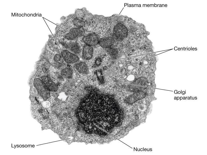
Animal Cell Electron Microscope Labelled. Typical animal cell pinocytotic vesicle lysosome golgi vesicles golgi vesicles rough er (endoplasmic reticulum) smooth er (no ribosomes) cell (plasma) membrane mitochondrion golgi apparatus nucleolus nucleus centrioles (2) each composed of 9. A typical animal cell (as seen in an electron microscope) medical images for powerpoint 1. The student concluded that the image of the plant cell obtained using the electron microscope. The diagram is very clear, and labeled;
 Images From qcpages.qc.cuny.edu
Cell is a tiny structure and functional unit of a living organism containing various parts known as organelles. 2.1 is an electron micrograph of part of an animal cell. But at the same time. (a) a mitochondria;b ribosomes (. (i) name the parts labelled as 1 to 10. See how a generalized structure of an animal cell and plant cell look with labeled diagrams.
The electron microscope two main advantages high resolving power (short wavelength of electrons) as electrons negatively are charged the beam can be focused 2.
One of the most elaborate duties that wellbeing and fitness experts face across their interaction with patients helps them recognise the issues and how to inspire them in regards to. (a) release of energy, (b) protein synthesis, (c) transmission of hereditary characters from parents to. The plant cell as more rigid and stiff walls. (1) it was discovered and named by porter (1948). The diagram is very clear, and labeled; Under the microscope priya observes a cell that has a cell wall and a distinct nucleus.
 Source: uni-mainz.de
All eukaryotic cells have a nucleus a plasma membrane cytoplasm endoplasmic reticulum ribosome s and golgi apparatus. Animal cells also have a because only plant cells perform photosynthesis, chloroplasts are found only in plant cells. Learn the structure of animal cell and plant cell under light microscope. The student concluded that the image of the plant cell obtained using the electron microscope. The electron microscope two main advantages high resolving power (short wavelength of electrons) as electrons negatively are charged the beam can be focused 2.
 Source: embryology.med.unsw.edu.au
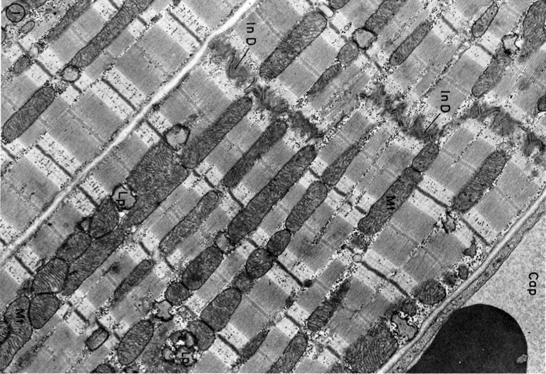
(b) idea of sections or cuts;idea of mitochondria orientated differently or in different positions / description of 3d structure of mitochondria, e.g. 560 x 364 pixel electron microscope image animal cell and organelles labeled animal cell plasma membrane organelles One of the most elaborate duties that wellbeing and fitness experts face across their interaction with patients helps them recognise the issues and how to inspire them in regards to. Label plant and animal cell labelled diagram. (a) a mitochondria;b ribosomes (.
 Source: uni-mainz.de
1.1 shows part of an animal cell viewed with an electron microscope. A typical animal cell (as seen in an electron microscope) medical images for powerpoint 1. State the advantages of using. Observe the labelled diagram of plant cell structure as given below: The plant cell as more rigid and stiff walls.
 Source: qcpages.qc.cuny.edu
560 x 364 pixel electron microscope image animal cell and organelles labeled animal cell plasma membrane organelles Animal cell diagram simple gcse. 1.1 (a) name the structures. Light microscope and the electron microscope. Animal cell (as seen under electron microscope).
 Source: labels-top.com
1.1 shows part of an animal cell viewed with an electron microscope. 560 x 364 pixel electron microscope image animal cell and organelles labeled animal cell plasma membrane organelles 2.1 is an electron micrograph of part of an animal cell. 80.the diagram below is a drawing of an organelle from a ciliated cell as seen with an electron microscope. The electron micrograph displayed below illustrates many of the plant cell characteristics discussed the cell wall, large central vacuole and chloroplasts are clearly visible also visible is the clearly defined nucleus containing chromatin nucleus chromatin the vacuole in this mature plant cell from a leaf is large, and occupies about 80% of
 Source: muskel-dnpb.blogspot.com
Under the microscope an animal cell shows many different parts called organelles that work together to keep the cell. State the advantages of using. The electron microscope two main advantages high resolving power (short wavelength of electrons) as electrons negatively are charged the beam can be focused 2. 1.1 (a) name the structures. Animal cells also have a because only plant cells perform photosynthesis, chloroplasts are found only in plant cells.
This site is an open community for users to do sharing their favorite wallpapers on the internet, all images or pictures in this website are for personal wallpaper use only, it is stricly prohibited to use this wallpaper for commercial purposes, if you are the author and find this image is shared without your permission, please kindly raise a DMCA report to Us.
If you find this site adventageous, please support us by sharing this posts to your preference social media accounts like Facebook, Instagram and so on or you can also save this blog page with the title animal cell electron microscope labelled by using Ctrl + D for devices a laptop with a Windows operating system or Command + D for laptops with an Apple operating system. If you use a smartphone, you can also use the drawer menu of the browser you are using. Whether it’s a Windows, Mac, iOS or Android operating system, you will still be able to bookmark this website.
Category
Related By Category
- Animal magic information
- Animal free shoes information
- Amazon prime anime information
- Anime awards 2017 information
- Animal crossing amiibo cards new horizons information
- Animal with i information
- 3d animation art styles information
- Animal crossing mole information
- Animated shakespeare information
- Animal kingdom tnt wiki information